Article No. VK-19010
Ultrasonic Imaging (Ultrasound Experimental Set 10)
Examining the possibilities and limits of the B-scan method with vivid tracking of the individual steps from a single ultrasound signal to the complete B-scan image
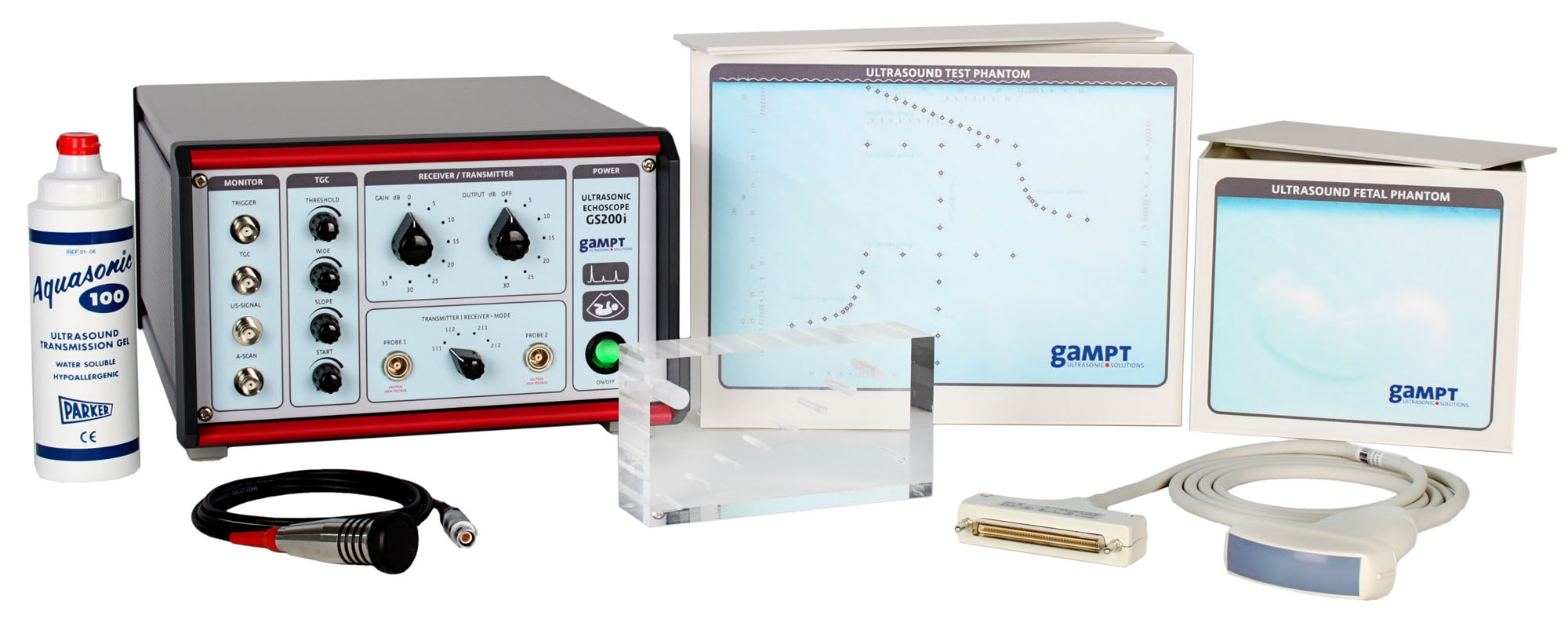
B-mode imaging is an ultrasound method frequently applied in medicine or in non-destructive material testing. Similar to X-ray or MRI procedures, the B-mode method delivers sectional images of the internal structure of a technical body or an organism, however without exposing it to radiation.This experimetal set was composed to be able to track the individual steps from the ultrasound signal to the complete B-scan image and to examine the possibilities and limits of the B-scan method as well as training its practical application. The set enables basic and application experiments for training and lab courses in medical and medical engineering specialities.The 2 MHz ultrasonic probe and the test block enable experiments in view of the fundamental physical principles of ultrasound propagation (time of flight, acoustic attenuation, reflection at boundaries, acoustic shadow, …). By using a single-element-transducer, it is possible to track the way from the ultrasound signal via the amplitude signal (A-scan), its conversion into a grey scale or coulor coded line scan and the composition of such line scans in a complete sectional image (B-scan).
For practical experiments, the set comprises two ultrasound phantoms with acoustic characteristics similar to those of human tissue.In order to image the internal structures of the phantoms, an array probe is used, as for example applied in medicine for examining the abdomen. This ultrasound probe possesses an array of 64 convexly arranged transducer elements. For the controlling of the array probe and the recording and analysis of the signal, a separate extension module is integrated in the GS200i ultrasonic echoscope.
The internal structures can be imaged and their dimensions can be measured by means of the measurement software. Furthermore, the influence of various parameters (focus, dynamic range, graphic filters, brightness, contrast, …) on the processing of signal and image can be examined.
| Ord.no. | Description |
|---|---|
| 10410 | Ultrasonic echoscope GS200i (incl. array probe) |
| 10152 | Ultrasonic probe 2 MHz |
| 10201 | Test block (transparent) |
| 10420 | Ultrasound test phantom |
| 10430 | Ultrasound fetal phantom |
| 70200 | Ultrasonic gel |
| Ord.no. | Description | for Experiment | |
|---|---|---|---|
| 10151 10154 |
Ultrasonic probe 1 MHz Ultrasonic probe 4 MHz |
PHY06 | Frequency dependence of resolution power |
| 10208 | Acoustic impedance samples | PHY21 | Reflection and transmission at boundaries |
| 10220 10154 |
Heart model Ultrasonic probe 4 MHz |
MED01 | Ultrasonic imaging at breast phantom |
| 10221 10151 |
Breast phantom Ultrasonic probe 1 MHz |
MED02 | Ultrasonic imaging at breast phantom |
| 10222 | Eye phantom | MED04 | Biometry at the eye phantom |
| 10224 10225 |
Ultrasound Breast Model with Cysts Ultrasound Breast Model with Malignant Tumors |
MED09 | Mamma sonography |
| 10440 | Ultrasound Gallbladder Model | MED10 | Gallbladder ultrasound |
