Article No. VK-PHY08
PHY08 Ultrasonic B-Scan
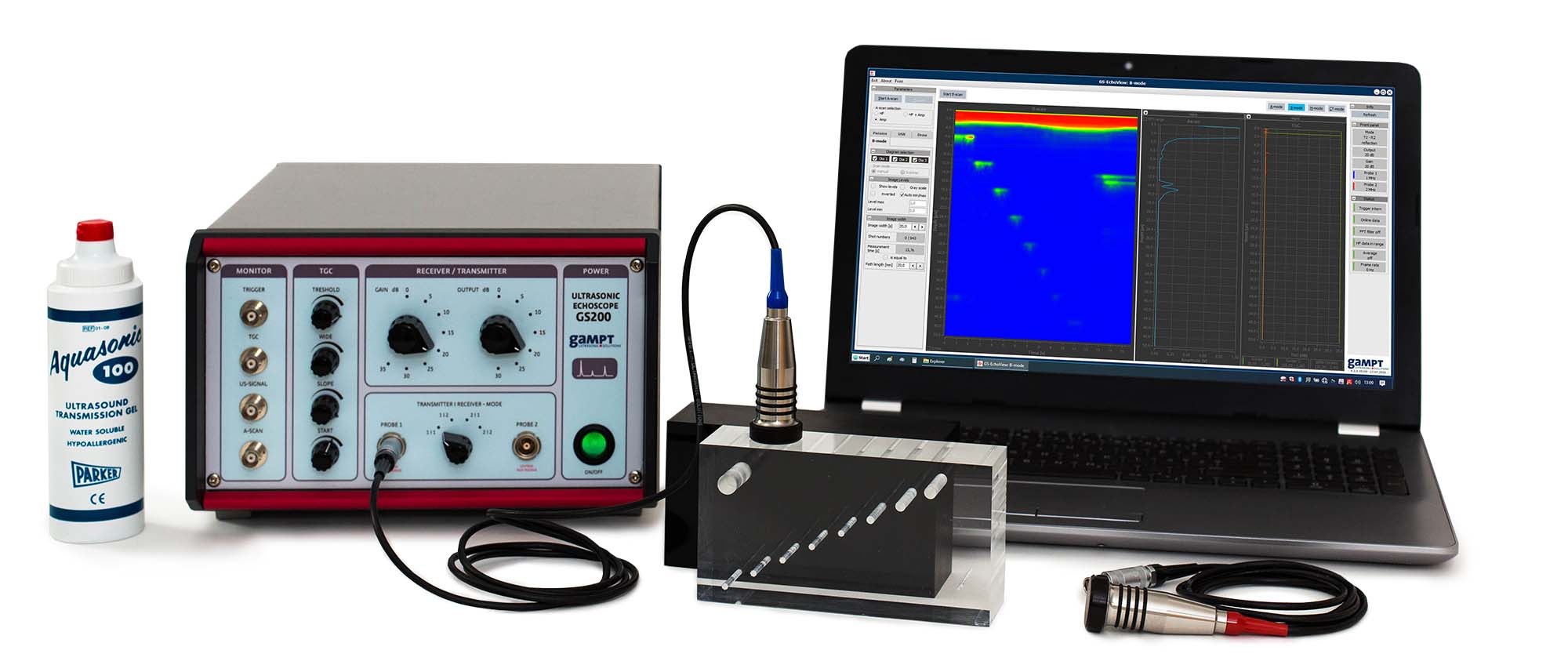
Demonstration of the basics of the B-scan method by recording the ultrasonic cross-sectional image of a simple test object "by hand" using an ultrasonic echoscope
By recording the ultrasonic cross-sectional image of a simple test object “by hand” using an ultrasonic echoscope, the basics of the B-Scan method are clearly demonstrated. Special features regarding scan quality such as sound focus, spatial resolution or imaging errors are investigated and analysed.
Keywords: Sound velocity, reflection, transmission, reflection coefficient, ultrasonic echography, A-Scan image, grey scale representation, B-Scan image, lateral resolution, focus zone, image artefacts
The conversion of the amplitude values of an amplitude scan into grey scale or colour values and the presentation of the time of flight as penetration depth yield a line of points with different brightness and/or colour values. The stringing together of such adjoining depth scans of an ultrasonic probe, which is guided along a line over the test area produces a sectional image, the so-called B-Scan image. Localisation along this line is based on the position of the probe and its movement speed. A simple way to obtain a B-Scan image is to guide the ultrasonic probe slowly by hand (compound scan). Scan quality is here dependent upon the coordinate-accurate transferral of scan points, the axial and lateral resolution of the ultrasonic probe, the grey scale and/or colour value resolution, the number of lines and imaging errors. In order to achieve e.g. exact lateral resolution, an additional coordinate-recording system is necessary such as a linear scanner.
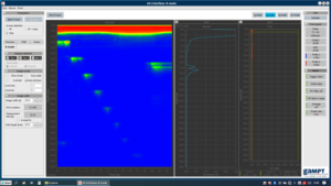
The screen shot of the measurement software shows the B-Scan presentation of the investigation of a sample with defined, built-in defects. The manual scanning of the block makes the functioning of the B-Scan method “easy to grasp”. The problems of movement artefacts with regard to lateral resolution caused “by hand” can be reduced by a mechanical scanning (PHY16).
| Ord.no. | Description |
|---|---|
| 10400 | Ultrasonic echoscope GS200 |
| 10151 | Ultrasonic probe 1 MHz |
| 10152 | Ultrasonic probe 2 MHz |
| 10201 | Test block (transparent) |
| 10204 | – optional: Test block (black) |
| 70200 | Ultrasonic gel |

