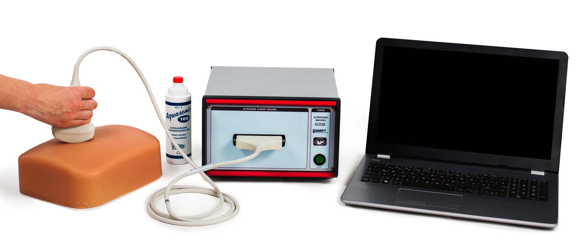
Article No. VK-MED10
MED10 Gallbladder ultrasound
Examination of a gallbladder model with serveral pathologies using the ultrasound B-image method
- Subject matter of the experiment
- Theoretical and practical aspects of the experiment
- Equipment
- Related Experiments
Introduction to ultrasound imaging diagnostics based on the B-scan method by examining a gallbladder model with simulated disease images of the gallbladder.
Keywords: Ultrasound imaging, B-scan method, array probe, gallbladder, gallstones, gries deposits, artefacts
Ultrasound imaging based on the B-mode procedure is an important tool in medical diagnostics. Similar to the X-ray or MRI procedure, the B-mode procedure provides sectional images of the internal structure of an organism, but without exposing it to radiation.In the experiment, the possibilities and limits of the B-scan procedure can be examined and the basic handling of an ultrasound B-scan device can be trained.An ultrasound model consisting of a block with enclosed gall bladders serves as the object of examination. These simulate various gallbladder diseases such as gallstones, thickening of the bladder wall or gallbladder deposits.
| Art.no. | Description |
|---|---|
| 10412 | Ultrasonic B-scan Device Gi210 |
| 10440 | Ultrasound Gallbladder Model |
| 70200 | Ultrasonic Gel |
| PHY01 | Basics of pulse echo method (A-Scan) |
| PHY06 | Frequency dependence of resolution power |
| PHY08 | Ultrasonic B-Scan |
| MED07 | Ultrasonic-Test-Phantom |
| MED08 | Ultrasonic-Fetus-Phantom |
| MED09 | Mamma sonography |
