Article No. VK-MED07
MED07 Ultrasound test phantom
Examination of an ultrasound test phantom with a normal beam probe and/or an array probe. Assessment of accuracy and performance of the ultrasound system in terms of resolution, dead zone and further parameters
- Subject matter of the experiment
- Theoretical and practical aspects of the experiment
- Results
- Equipment
- Related Experiments
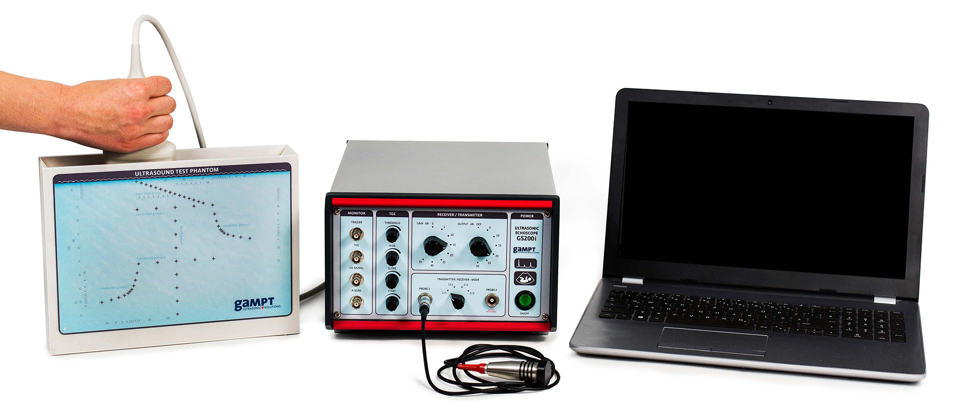
In the experiment, an ultrasound test phantom is examined with a normal beam probe and / or an array probe. On thebasis of bar-shaped targets, which are arranged in the form of functional groups, the accuracy and performance of theused ultrasound system is assessed in terms of resolution, dead zone and further parameters.
Keywords: Time of flight of sound, sound velocity, acoustic attenuation, sound field, reflection, A-scan, B-scan, grey tone coding, resolution, focusing, transmitting power, total gain, TGC, dynamic range, dead zone
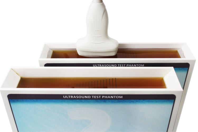
Ultrasonic test phantoms are used for quality assurance and routine check of the accuracy and performance of ultrasound imaging systems. Such ultrasound test phantoms are also excellent test objects for introduction to two-dimensional ultrasound imaging.The tissue-equivalent material of test phantoms for medical ultrasonic systems usually has physical characteristics which are similar to the acoustic characteristics of the human tissue. Various test structures can be embedded in the material which allow for the objective and comparable evaluation of the display characteristics of ultrasonic devices.
GS200i:
Using the the 2 MHz normal normal beam probe, A-scan and hand-guided B-scan images of the phantom are recorded. From time-of-flight measurements of the echoes of the vertical and horizontal test groups, an actual sound velocity in the phantom of approximately 1465 m/s is determined.
GS200i or Gi210:
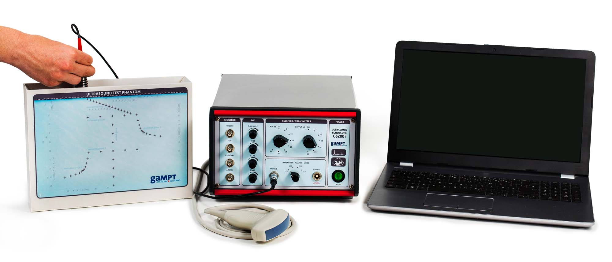
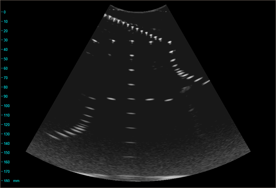
Axial distance measurements in the Bscan images taken with the array probe (left figure) show a measurement error of approximately 5% assuming a sound velocity of 1540 m/s. This value is an average of the velocities of sound in various human tissues and is used as the default in many medical ultrasound systems.The investigation of the axial and lateral resolution test groups (right figure) shows improvements in the resolution of the measurement system as the frequency of the sound increases, and its deterioration as the depth of measurement increases.
| Art.No. | Description |
|---|---|
| 10410 | Ultrasonic echoscope GS200i |
| or | |
| 10412 | Ultrasonic B-scan device Gi210 |
| 10152 | Ultrasonic probe 4 MHz |
| 10420 | Ultrasound test phantom |
| PHY01 | Basics of pulse echo method (A-scan) |
| PHY08 | Ultrasound B-scan |
| MED08 | Ultrasound fetal phantom |
| MED09 | Mamma sonography |
| MED10 | Gallbladder ultrasound |


